People who have progressed to central vision loss from any one of a number of retinal dystrophies have probably noticed a tiny spot of healthier sight in the very center of the macula (the fovea).
Over time, the fovea becomes surrounded by a growing band of blindness, first appearing as individual spots, then coalescing to resemble a doughnut with a very small hole in the middle. This is the final bunch of cells to atrophy, usually long after the surrounding tissue has done so. The process is known as “foveal sparing”. With a diameter of only about 1.5 millimeter, the spared fovea is sometimes good enough to help identify a single letter on a doctor’s eye chart, but it is usually not wide enough to be functionally usable.
So what natural process is protecting the fovea? Even more thought-provoking, if the process is identified, could it be duplicated and applied to the outlying rod and cone photoreceptor cells in the macula? Retinal dystrophies that might benefit include Stargardt disease, pseudo-Stargardt pattern dystrophy, central areolar choroidal dystrophy, diabetic retinopathy, mitochondrial retinal dystrophy, and geographic atrophy (dry age-related macular degeneration). The cause of foveal sparing is not yet completely known, but a study* published in IOVS in August 2019 offers seven reasonable hypotheses:
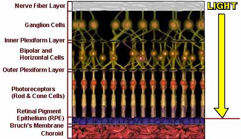
1. Cone cells are responsible for central, bright light, fine detail and color vision. They are most abundant in the macula. Rod cells are responsible for peripheral and dim light vision. A substance known as rod-derived cone viability factor (RdCVF) is secreted from rod cells that surround the fovea and protect the cone cells from degeneration. It has been proposed that the cone cells in the fovea are either more sensitive to the RdCVF, or the level of secretions in the fovea is higher than in the the surrounding macula.
2. The natural carotenoids lutein, zeaxanthin, and meso-zeaxanthin are responsible for the yellow color (pigment) of the macula, which protects the cells by filtering blue light. These carotenoids may be more concentrated in the fovea.
3. The density of the cone cells is much higher in the fovea than in the surrounding macula. In other words, the fovea has a much larger store of cone cells, which takes longer to destroy.
4. Neither rod cells nor S-cone cells (the ones responsible for perceiving blue) exist in the fovea. These cells are susceptible to aging and disease, so their absence may be helping the fovea to remain healthier longer.
5. Retinal pigment epithelial (RPE) cells supply nutrition to the photoreceptor rods and cones. There is an unfavorably high ratio of rods per RPE cell in the retina outside of the fovea, which could explain why the fovea remains healthier longer than the surrounding macula
6. The unique blood supply to the foveal area (no blood vessels) has been suggested as having a protective effect.
7. Genetic variances may influence the durability of the fovea.
These are possible explanations for foveal sparing as outlined by the study’s research team. As for duplicating the body’s natural protections of the fovea to benefit the surrounding macula, the team concluded that “Further research is necessary to elucidate the mechanisms…The new insights gleaned from these studies might pave the way for new disease-independent [gene therapies] for prolonged preservation of foveal tissue in degenerative retinal disease”.*
*Investigative Ophthalmology & Visual Science. Nathalie M. Bax, et al (August 2019, Vol.60, 3414-3420. doi:10.1167/iovs.19-28020)